The Step-by-Step Guide to Zygomatic Surgery
July 24, 2026
What Is Zygomatic Implant Surgery — and Is It Right for You?
Zygomatic implant surgery is a specialized procedure that anchors long titanium implants into the cheekbone (zygoma) rather than the upper jawbone — making it possible to place a full fixed set of teeth even when the jaw has little to no usable bone.
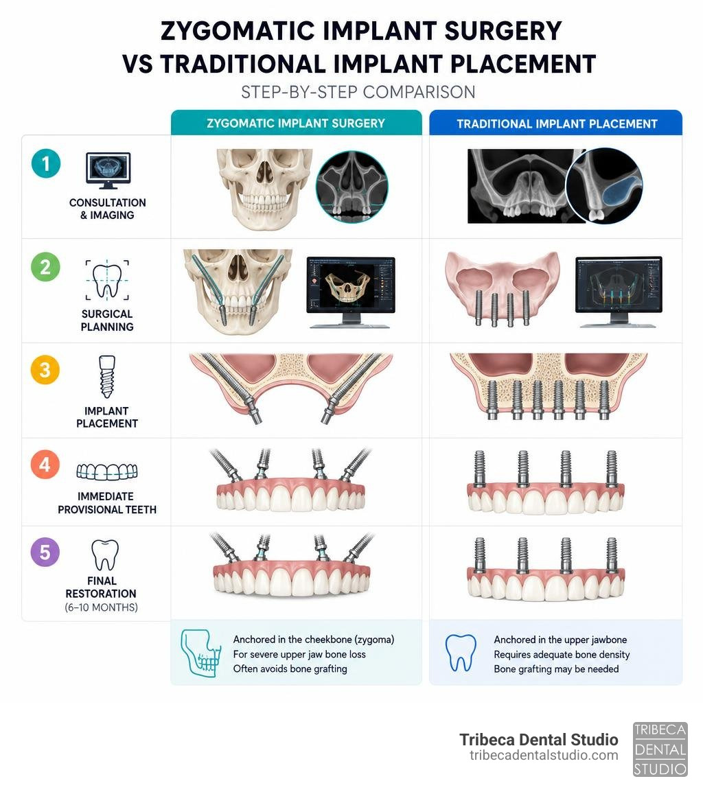
Here is a quick overview of how the process works:
- Consultation & imaging — A CBCT scan maps your bone anatomy and sinus health.
- Surgical planning — Implant length, angle, and placement path are planned in 3D software.
- Implant placement — Long implants (typically 30–52 mm) are placed into the zygomatic bone at a precise angle, bypassing the atrophied upper jaw entirely.
- Immediate provisional teeth — In most cases, a fixed temporary bridge is attached the same day.
- Final restoration — After 6–10 months of healing, a permanent zirconia bridge replaces the provisional.
This approach is specifically designed for patients with severe upper jaw bone loss — people who have often been told they are not candidates for traditional implants and who want to avoid lengthy bone grafting procedures.
It is a technically demanding surgery. The implants are significantly longer than standard implants, placed at a steep angle, and anchored in a completely different part of the skull. Done well, the results can be life-changing. Done poorly, the risks — including sinus complications and implant failure — are real.
This guide walks you through every step, from candidacy to final prosthesis.
I’m Dr. Nina Izhaky, a graduate of New York University College of Dentistry and founder of Tribeca Dental Studio, where I work alongside an on-site team of specialists — including oral surgeons — to help patients navigate advanced solutions like zygomatic implant surgery. My goal with this guide is to give you a clear, honest picture of what the procedure involves so you can make a confident, informed decision.
Discover more about zygomatic implant surgery:
Understanding Zygomatic Implant Surgery: Indications and Candidacy
When you lose teeth, the alveolar bone—the portion of the jawbone that holds your tooth roots—begins to resorb. Over time, or due to severe periodontal disease, facial trauma, or long-term denture wear, this bone loss can become so extreme that traditional dental implants simply have nothing to hold onto.
Historically, patients in this situation had to undergo aggressive bone grafting procedures (such as taking bone from the hip or tibia) followed by months of waiting. Today, zygomatic implant surgery offers a highly successful, graftless alternative. By utilizing the dense, stable bone of the zygomatic arch (your cheekbone), we can secure a stable foundation for a full-arch restoration.
To understand where this treatment fits in, let us compare the primary restorative paths for severe bone loss:
| Feature | Traditional Dental Implants | Advanced Bone Grafting + Implants | Zygomatic Implant Surgery |
|---|---|---|---|
| Primary Anchorage Site | Alveolar jawbone (maxilla) | Grafted bone in the jaw | Zygomatic bone (cheekbone) |
| Need for Bone Grafting | None to minimal | Extensive (sinus lifts, block grafts) | None (graftless protocol) |
| Treatment Timeline | 3 to 6 months | 9 to 18+ months (multiple stages) | Same-day provisional teeth |
| Surgical Complexity | Standard | Moderate to High | Advanced (requires specialist) |
| Long-Term Success Rate | ~95% | ~90% | ~95% to 98% |
To evaluate the degree of jawbone shrinkage, specialists utilize the Cawood and Howell classification system. Patients who fall into Class V (flat ridge with loss of alveolar height and width) and Class VI (depressed ridge with basal bone loss) are the prime candidates for zygomatic solutions.
If you are trying to weigh your options, you can read more about why this approach is often preferred in our article on Why Zygomatic Implants Beat Bone Grafting Every Time. Furthermore, we break down how modern technology allows us to bypass traditional preparatory surgeries in our guide to Can You Get Dental Implants Without a Bone Graft?.
Ideal Candidates and Contraindications
While zygomatic implants are a rescue therapy, they are not suitable for everyone.
Ideal candidates include:
- Patients with severe maxillary bone resorption (Cawood and Howell Class V or VI).
- Individuals who have experienced failed bone grafting or failed sinus lifts.
- Patients who have lost conventional implants and need a reliable “rescue” option.
- Maxillofacial reconstructive patients who have suffered tumor resections, cleft palate, or severe facial trauma.
Contraindications and Ineligibility Factors: According to clinical standards outlined in the ITI consensus report on zygomatic implants: indications, evaluation of surgical techniques and long-term treatment outcomes, certain conditions may make a patient ineligible for this procedure.
Absolute contraindications include active, untreated maxillary sinus pathology (such as severe acute sinusitis), history of head and neck radiation therapy targeting the midface, and uncontrolled systemic metabolic diseases like uncontrolled diabetes. Relative contraindications include heavy smoking, active periodontal disease in remaining lower teeth, and chronic steroid or bisphosphonate therapy, which can impair bone healing. Any active sinus infection must be fully treated and resolved before we can proceed with surgical placement.
Preparing for Zygomatic Implant Surgery: Preoperative Evaluation
A successful surgical outcome depends entirely on thorough preoperative planning. Because the surgical path runs close to vital anatomical structures—including the maxillary sinus, the infraorbital nerve, and the orbit of the eye—we leave absolutely nothing to chance.
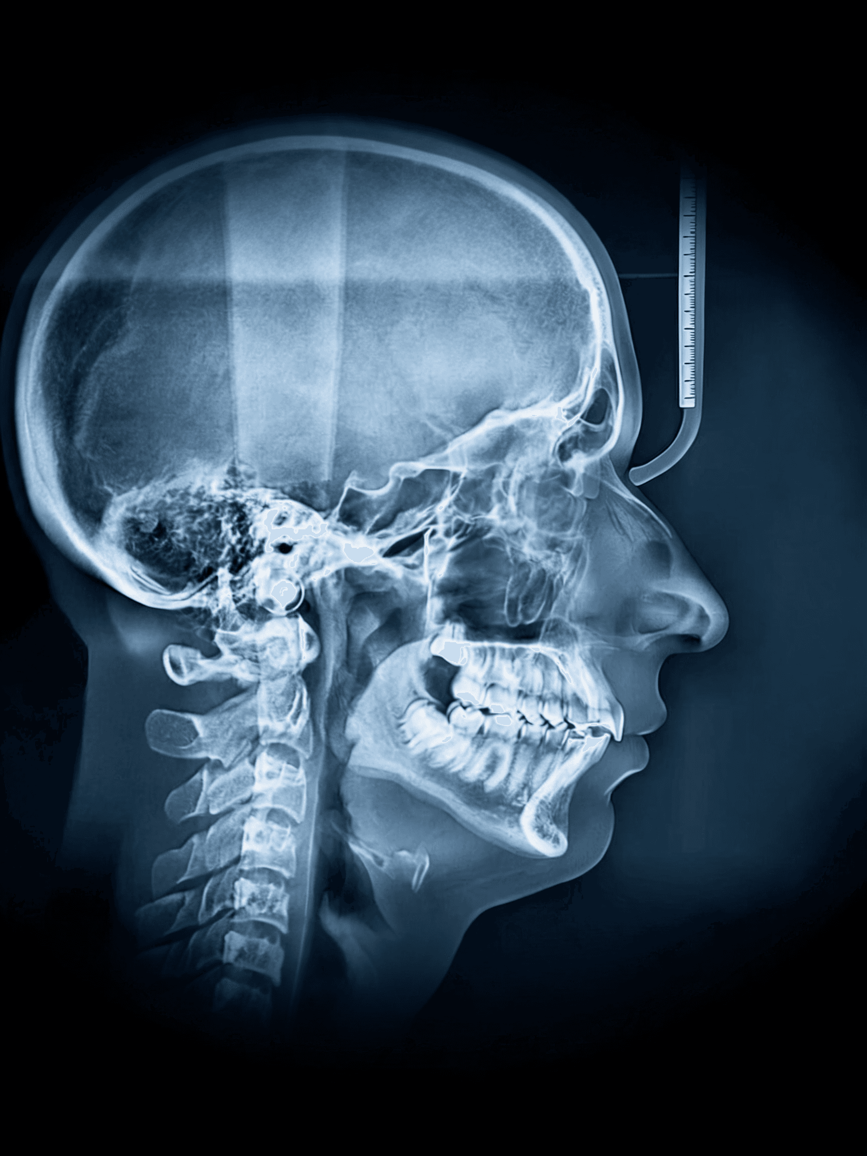
The cornerstone of this planning is Cone Beam Computed Tomography (CBCT) imaging. This 3D imaging allows us to:
- Assess the volume, density, and three-dimensional shape of the zygomatic bone.
- Evaluate the health of the maxillary sinuses, looking for signs of chronic sinusitis, polyps, or mucosal thickening.
- Identify the exact path of the infraorbital nerve and the lower borders of the orbit to prevent injury.
- Perform virtual 3D planning using specialized software to choose the exact length, angulation, and exit point of each implant before the patient ever enters the surgical suite.
For a deeper look into how we prepare our patients for this transformative journey, check out our comprehensive Zygomatic Dental Implants Guide 2026.
Surgical Techniques: Intrasinus vs. Extrasinus Approaches
Over the last few decades, the surgical protocols for placing zygomatic implants have evolved significantly to improve patient comfort, optimize the position of the teeth, and minimize sinus-related side effects.
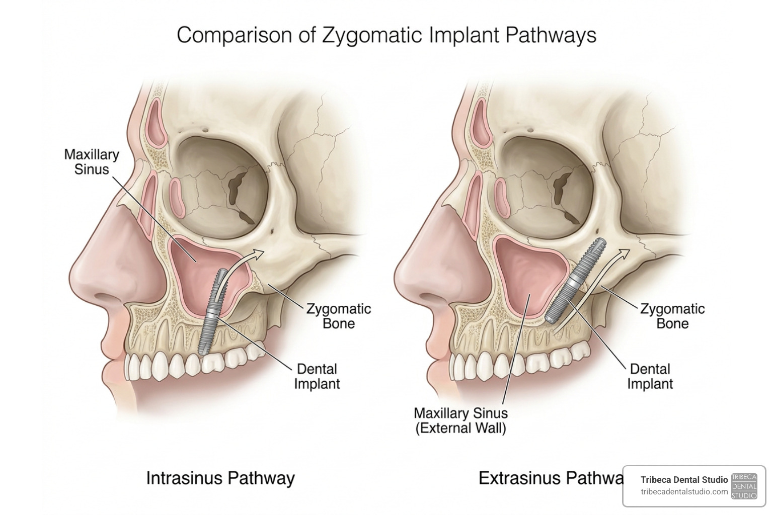
Historically, the original technique developed by Professor Per-Ingvar Brånemark in the late 1980s utilized an intrasinus approach. Over time, alternative protocols like the sinus slot technique and the extrasinus (extramaxillary) protocol emerged. Today, the gold standard for personalizing this procedure is the Zygomatic Anatomy Guided Approach (ZAGA) classification, which categorizes a patient’s maxillary wall anatomy into five distinct types (Type 0 to Type IV) to guide the surgeon’s path.
To understand these developments, you can read the detailed clinical analysis in The zygoma anatomy-guided approach (ZAGA) for rehabilitation of the atrophic maxilla.
Intrasinus Technique Advantages and Risks
The traditional intrasinus technique involves creating a small window in the lateral wall of the maxillary sinus, gently elevating the delicate sinus lining (the Schneiderian membrane), and passing the implant directly through the sinus cavity to reach the cheekbone.
- Advantages: It provides a completely internal pathway, keeping the implant body covered by the lateral maxillary wall.
- Risks: Because the implant spends a large portion of its length inside the sinus cavity, this technique carries a higher risk of post-operative sinus issues. If the Schneiderian membrane is compromised or if the implant does not seal properly at the entry point, bacteria can enter the sinus cavity. This can lead to chronic sinusitis or mucosal thickening. In fact, clinical studies show that asymptomatic mucosal thickening on imaging studies is present in approximately 20% of patients after zygomatic implant placement.
Extrasinus and ZAGA Protocols
To address the prosthetic and biological limitations of the intrasinus approach, the extrasinus (or extramaxillary) protocol was developed. Instead of drilling through the sinus, the implant is placed along the outer curvature of the maxillary wall, bypassing the sinus cavity entirely or only touching its outer border.
- Advantages: This technique minimizes sinus membrane disruption, reducing the risk of chronic sinus infections. Furthermore, because the implant is positioned more toward the cheek side, the prosthetic platform emerges closer to the top of the dental ridge (buccal emergence) rather than the roof of the mouth. This makes the final bridge much less bulky, easier to clean, and far more comfortable for speaking and chewing.
- Specialized Designs: Advanced systems utilize specialized implants like ZAGA Flat implants, which feature a flattened, unthreaded middle section that rests smoothly against the outer maxillary wall, preventing soft tissue recession and irritation.
For patients seeking highly refined, minimally invasive options, techniques like the Parisi-Févola Technique: Minimally Invasive Protocol for Zygomatic Implant Placement Under Local Anesthesia demonstrate how modern extrasinus approaches can even be performed comfortably in an outpatient setting under local anesthesia.
The Step-by-Step Surgical Procedure
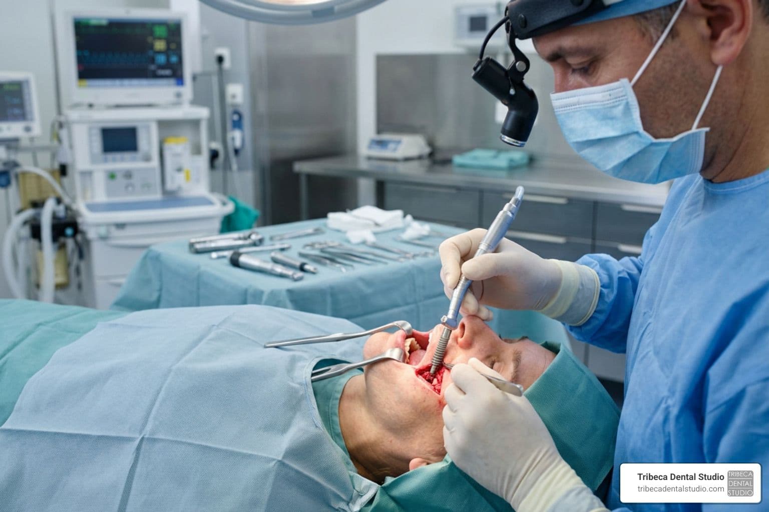
Placing zygomatic implants requires an extraordinary level of surgical precision and specialized instrumentation.
Because these implants must traverse a long distance from the mouth to the cheekbone, they are significantly longer than standard dental implants. Standard implants are typically 8 to 14 mm long; zygomatic implants range from 30 mm to 60 mm in length.
To achieve successful osseointegration, the implant must achieve high primary stability (mechanical tightness in the bone) upon insertion. We measure this using insertion torque, aiming for a minimum of 35 to 50 Ncm (Newton-centimeters) of torque, which allows us to safely attach temporary teeth on the very same day.
Step-by-Step Protocol of Zygomatic Implant Surgery
Here is a detailed, chronological breakdown of how our surgical specialists perform this advanced procedure:
Step 1: Anesthesia and Patient Comfort
The procedure begins by ensuring the patient is completely comfortable and pain-free. While many cases are performed under general anesthesia or deep IV sedation, experienced surgeons can also perform the surgery under local anesthesia. We administer highly targeted nerve blocks to numb the infraorbital, greater palatine, nasopalatine, and superior alveolar nerves.
Step 2: Incision and Flap Elevation
A crestal incision is made along the upper ridge, with releasing incisions to allow the surgeon to raise a full-thickness mucoperiosteal flap. This flap is carefully elevated to expose the lateral wall of the maxilla, the zygomatic buttress, and the lower border of the orbital rim. Direct visualization of these landmarks is critical to ensure safety and prevent disorientation during drilling.
Step 3: Preparing the Implant Site (Osteotomy)
Using specialized, extra-long zygomatic drills and protective drill guards to protect the surrounding soft tissues, the surgeon begins preparing the channel.
- A round bur or needle drill is used to mark the entry point on the alveolar ridge.
- The drill is angled at approximately 45 to 60 degrees, pointing toward the body of the zygoma.
- Sequential drilling is performed at a controlled speed (usually 1,000 to 1,500 rpm) with continuous sterile saline irrigation to prevent heat damage to the bone.
Step 4: Verifying Depth and Path
Once the osteotomy is complete, the surgeon uses a specialized depth gauge to measure the exact length of the channel, ensuring that at least 7 mm of the implant tip will be firmly anchored within the dense cortical bone of the cheekbone.
Step 5: Implant Insertion
The selected zygomatic implant is carefully threaded into the prepared channel. It is driven in using a handpiece and completed with a manual torque wrench to monitor the exact insertion torque. We aim for a high insertion torque (up to 50 Ncm) to ensure the implant is locked solid.
Step 6: Abutment Connection and Closure
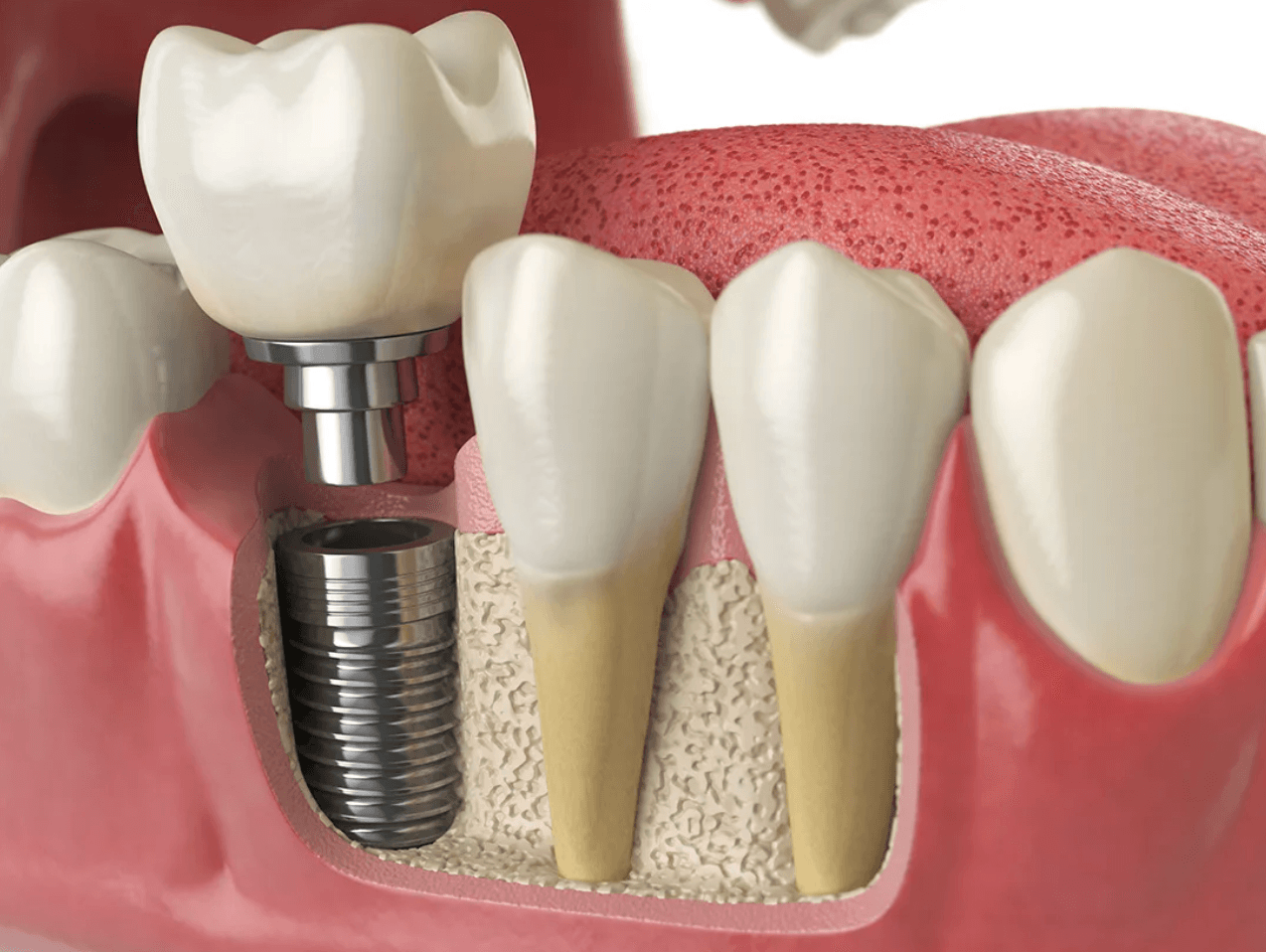
Once the implants are securely in place, angled Multi-Unit Abutments (ranging from 17° to 60°) are screwed onto the implant heads. These angled abutments correct the steep entry angle, ensuring that the screw holes for your new teeth exit straight up through the chewing surfaces rather than pointing out toward your lips. The gum tissues are then closed and sutured with precision.
Our surgical team, led by highly trained specialists, coordinates every step of this delicate process. You can learn more about our surgical expertise by visiting the profile of our Dental Implants Board Certified Implant Specialist Dr. Sam.
Postoperative Care, Immediate Loading, and Long-Term Outcomes
One of the greatest benefits of modern zygomatic implantology is the ability to walk out of the clinic with a functional, beautiful smile on the very same day.
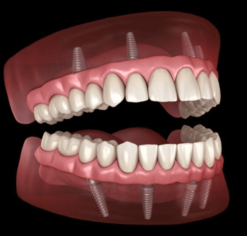
This process is known as immediate loading or immediate provisionalization. When we achieve excellent primary stability during surgery, we can immediately attach a provisional PMMA (acrylic) bridge. This temporary bridge is screw-retained and acts as a splint, connecting all the implants together. This “cross-arch stabilization” distributes chewing forces evenly, protecting the implants while they fuse with your bone.
You will wear this provisional bridge for approximately 6 to 10 months. Once osseointegration is complete, we capture digital impressions to design and manufacture your final, ultra-strong zirconia bridge, which is custom-milled for optimal aesthetics, strength, and hygiene.
To explore our full range of implant options and prosthetic materials, visit our dedicated page on Services: Dental Implants.
Managing Complications and Long-Term Success
Zygomatic implants have a phenomenal track record. Clinical literature shows that the 12-year survival rate of zygomatic implants is approximately 95%, with some studies reporting success rates up to 98% when proper protocols are followed.
However, as with any advanced surgical procedure, complications can occur.
Common Potential Complications:
- Sinusitis: This is the most common complication, affecting roughly 14.2% of patients over the long term. It is often managed successfully with antibiotics and decongestants, though severe cases may require sinus lavage or conservative surgical intervention.
- Asymptomatic Mucosal Thickening: Seen in about 20% of patients on post-operative CBCT scans, this thickening of the sinus lining is usually harmless and requires no active treatment unless symptoms develop.
- Local Soft Tissue Issues: Minor gum recession or exposure of the implant neck can occur, particularly with older intrasinus techniques. This is minimized today by utilizing extrasinus approaches and protective tissue flaps (like the cheek fat pad transposition).
- Temporary Paresthesia: Slight numbness in the cheek or lip can occur if the surrounding nerves are stretched during surgery, but this is almost always temporary and resolves on its own.
To learn more about how we prevent, identify, and manage these issues, read our detailed guide on Zygomatic Dental Implants Complications.
Frequently Asked Questions about Zygomatic Implants
Can zygomatic implants be placed under local anesthesia?
Yes, they absolutely can. While general anesthesia or deep IV sedation is traditional for extensive midface surgeries, modern minimally invasive protocols have made local anesthesia a highly predictable and comfortable option.
Using the Parisi-Févola Technique: Minimally Invasive Protocol for Zygomatic Implant Placement Under Local Anesthesia, surgeons utilize a small 1.5 cm incision and minimal flap elevation to place the implants. Performing the procedure in an outpatient setting under local anesthesia reduces post-operative swelling, eliminates the risks and recovery time associated with general anesthesia, and allows for active patient collaboration during the procedure.
How do zygomatic implants compare to pterygoid implants?
Both zygomatic and pterygoid implants are “graftless” solutions designed to bypass severe maxillary bone loss, but they utilize different anchorage sites in the skull.
- Zygomatic Implants: Anchor into the cheekbone. They are ideal for patients with bone loss in both the front (anterior) and back (posterior) of the upper jaw.
- Pterygoid Implants: Anchor into the pterygoid plate of the sphenoid bone, located at the very back of the upper jaw. They provide crucial posterior support, allowing us to avoid sinus lifts and extend the dental bridge all the way back to the molars.
Often, we combine these two approaches (using zygomatic implants in the front/middle and pterygoid implants in the back) to achieve maximum stability without any bone grafts. To see a detailed head-to-head comparison, check out our guide on Zygomatic vs Pterygoid: Unpacking Advanced Dental Implant Options.
What is the recovery timeline after zygomatic surgery?
While the thought of cheekbone implants sounds intense, most patients are pleasantly surprised by how manageable the recovery is.
- First 2 to 3 Days: You can expect some swelling and mild bruising around the cheeks and under the eyes, along with minor nosebleeds (epistaxis). This is completely normal and easily managed with ice packs and prescribed pain relievers.
- First 2 Weeks: Initial soft tissue healing takes place. You will need to maintain a soft-food diet to avoid putting excessive force on the healing implants.
- 3 to 6 Months: This is the critical window for osseointegration, during which your cheekbone fuses permanently with the titanium implant body. You will continue to wear your provisional bridge during this time.
To help you prepare for every stage of your post-operative journey, we have mapped out a detailed recovery road map in Your Timeline for Zygomatic Implant Healing.
Conclusion
Severe bone loss in the upper jaw does not mean you are destined to wear loose, uncomfortable dentures for the rest of your life. Thanks to the predictable, long-term success of zygomatic implant surgery, we can bypass the jawbone entirely to anchor a beautiful, permanent smile directly into the stable bone of your cheeks.
Because this surgery is highly technique-sensitive, choosing the right team is paramount. At Tribeca Dental Studio in New York City, our founder, Dr. Nina Izhaky, has built a world-class, multi-specialty practice where prosthodontists, general dentists, and board-certified oral surgeons work side-by-side. This collaborative, in-house approach ensures that your surgical placement is planned in perfect harmony with your final cosmetic results.
If you have been told you are not a candidate for dental implants, or if you want to avoid the long, painful road of bone grafting, we invite you to experience our personalized, family-centered care.
Ready to take the first step toward a confident, permanent smile? Schedule your personalized consultation today by visiting our Services: Zygomatic Implants page.